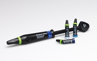
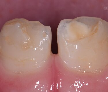
A healthy 11 year old male presented on referral to the practice with a chief concern of unstable and irregular composite restorations affecting his maxillary central incisors. The teeth were initially described in a classic molar-incisor hypomineralization pattern with an uncavitated, brown hypomineralization lesion localized to tooth 11 and tooth 21MI status post uncomplicated enamel fracture secondary to impact with a metal drink bottle (CPT_2084). The patient was described as being in a mid-mixed dentition state. Esthetic treatment options were outlined including the possibility of pre-prosthetic whitening, which was refused by the parents after learning that further whitening procedures would need to likely be completed in the future as the remaining secondary teeth erupted intraorally, with the risk of color variance with the bleached teeth. Whitening acts to decrease the chromatic aspects of the hypomineralized lesions and simultaneously lifts the value of the background shade, decreasing the visual contrast between lesion and tooth. Resin infiltration is always an option for uncavitated hypomineralized lesions, however, with residual composite covering the teeth, structural deficits from trauma and the presence of chromatic regions within the area of organic rich hypomineraization, a conservative reductive approach was elected, both to increase the predictability of bonding in the region and to visually eliminate the lesion of interest.
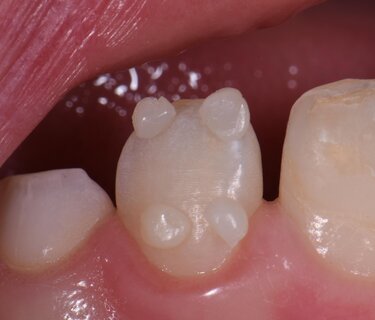
Shade selection was completed prior to the application of an 18% benzocaine/2% tetracaine-based topical anesthetic (Zap, Germiphene, Brantford, ON, Canada). It is known that dehydration decreases the water content, which increases the proportionate amount of air in a tooth, decreasing the refractive index from 1.33 (water) to 1.00 (air) thereby increasing the reflective index and thus the visual value and opacity (CPT_2085). Empress Direct (Ivoclar Vivadent, Schaan) composite shade buttons were selected and placed overlapping the incisal region of tooth 22, which functioned as the color reference tooth. Corresponding dentin shades were placed cervically, where the enamel was thinnest and the dentin hue most appreciable. A marked halo and sub halo translucency was noted as part of the color map. An enamel shade of A1 and a dentin shade of A2 was selected for the case (CPT_2086).
Following topical anaesthesia application for 90 seconds and application of 1.4 carpules of a 2% Lignocaine with 1:100,000 epinephrine solution (Septodont) via buccal infiltration, the region was isolated with a split rubber dam with clamp anchors on the upper E’s (CPT_2087). The old restorative material was removed, and the hypomineralized region conservatively reduced to expose an improved amount of inorganic substrate for bonding. It was noted that there were hypomineralized regions in both the middle and incisal thirds of both 11 and 21. A partial thickness oblique fracture was noted in the enamel but did not penetrate to the palatal surface.
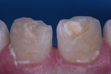
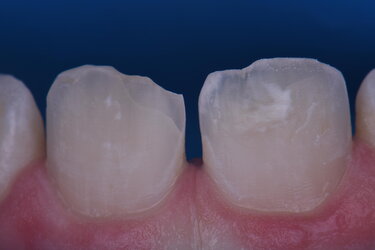
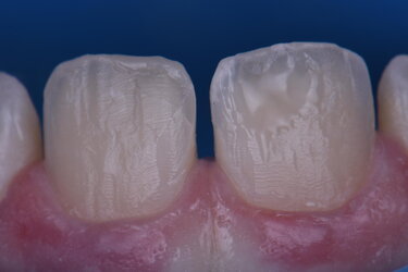
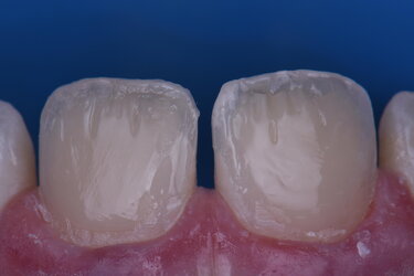
It was decided to leave this area and reinforce it with bonded restorative material in the spirit of minimal invasion (CPT_2088).
The surface was isolated for a total etch adhesive approach, and the first thin lingual shelf region sculpted and defined with shade A2 Enamel with a halo of A2 Dentin (Empress Direct, Ivoclar Vivadent). This area was finessed as thin as possible in order to allow room for subsequent layers which would define the desired translucency in this zone. Resin coating was achieved in the hypomineralized zone using 3 micro layers of A2 Tetric Evoflow (Ivoclar Vivadent) (CPT_2089).
The area of hypomineralization on both teeth were addressed with the next, thin A2 Dentin layer, which aimed to harmonise body value and chroma and create incisal reaching irregularities typical of internal dentin anatomy. Greater definition and correction of the halo effect value was desired and achieved using a custom mixture of Empress Color White : Ochre in a 9:1 ratio. This was delivered to the halo using a custom fissure sculpting instrument (TNTAM1, Hu-Friedy Corp, IL) (CPT_2090).
A degree of opalescence was desired between the incisal fingerlings, and thus applied using shade Opal Trans (Empress Direct, Ivoclar Vivadent) using an Optrasculpt instrument (Ivoclar). Of note, use of the Optrasculpt instrument obviates or minimises the clinical need to use dipping resin, which has the ability to weaken or alter the physical properties of composite if used in excessive quantities. Often, singular or multiple fine opaque connections to the dentin are seen from halo to dentin body internally. This was accomplished by using Empress Direct White and in this case is placed superficial to the cured Opal Trans layer (CPT2091).
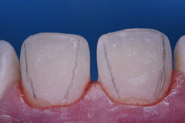
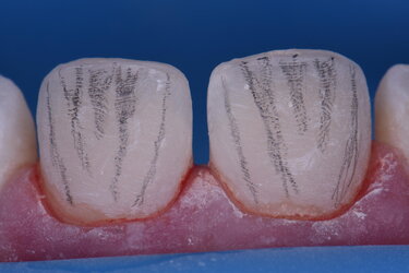
The final A1 enamel layer was sculpted in place (Empress Direct, Ivoclar Vivadent) using the Optrasculpt instrument, and primary anatomy shaped and defined (CPT_2092). Secondary anatomy was mapped and sculpted using a series of needle point and thin chamfer fine diamond burs (Mani) (CPT_2093)before final finishing and polishing using abrasive discs and the Optragloss two-step polishing system, which nicely highlighted the secondary and tertiary anatomy (CPT_2094).
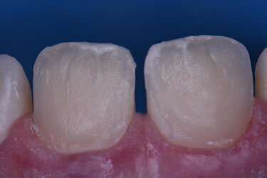
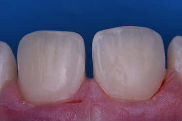
Post-operative analysis indicates successful visual elimination of the areas of concern, recreation of incisal maverick and translucency features, and offers a smooth, bio-anatomical surface that should rectify any concerns the patient has relative to smiling confidence for many years to come (CPT_2097).
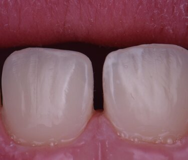
Incisor hypomineralization is a subset of molar-incisor hypomineralization, a condition which is esthetically and often functionally debilitating in affected individuals. It features a multifactorial etiology affecting approximately 16% of the Northern European population (Weerheijm, 2003) in low or under-fluoridated communities. The period of susceptibility is from 32 weeks in utero to 5.5 years of age and results in an enamel lesion defect that initiates from the level of the dentinoenamel junction (DEJ) and extends superficially. Fluorosis lesions in contrast are due to the presence of excessive systemic levels of fluoride and feature extension from the surface towards the DEJ. The defect of MIH or incisal hypomineralization is noted in the post-secretory stage of amelogenesis, which leaves a surface which is weak and susceptible to rapid post-eruptive breakdown from masticatory and environmental forces, increasing the risk of secondary decay. This resulting surface is defined as more irregular, porous with hydroxyapatite crystals unorganized, visually indistinct and deficient in volume. Crombie et al (2013) determined the inorganic content of affected enamel lesions as 58.8% (vol%) relative to unaffected enamel at 86%.
Strategies are graded from non-invasive to progressively more invasive depending on the desires of patient and guardian if applicable. Both hydrogen and carbamide peroxide-based whitening protocols have been used successfully with or without resin infiltration strategies, the latter having widely varying success in the literature (Kumar, 2017).
The patient in this case with the support of his mother decided that both pre-prosthetic whitening and resin infiltration were to have a limited cost:benefit ratio as other teeth yet to be erupted intraorally may feature a darker chroma or value and require subsequent whitening. As there was already a deficient volume of enamel due to trauma, paired with the prominent chromatic hypomineralized lesion in 11, the decision was made to proceed with a reductive approach alone. This allows simultaneous reduction in substrate high in organic content and increases the mineral density of the resulting substrate, simultaneously providing space for corrective resin layering with a more predictable adhesive shear bond strength (Fayle, 2003).
Empress Direct was chosen as a direct restorative material as it exhibits extremely tight tolerances with respect to translucency, opacity and fluorescence relative to nature. Barium glass diameters of 0.7 microns for dentin and 0.4 microns for enamel ensure clinical performance relative to strength and wear resistance as pertains to each layer. Polymerization shrinkage is controlled in the dentin layer which is often applied in a more generous application using pre-polymers, which simultaneously increase its strength. Radiopacity is boosted using ytterbium trifluoride, which also has fluoride release as an adjunct. It is a material designed to perform optically, clinically and functionally as optimally as possible using a resin composite enamel-dentin substitute and remains a gold standard in modern direct restorative armamentaria.
-
Crombie, F, Manton DJ, Palamara JEA, Zalizniak I, J Cochrane N, Reynolds E. 2013. Characterisation of developmentally hypomineralised human enamel. J Dent. 41. 10.1016/j.jdent.2013.05.002.
-
Fayle SA. 2003. Molar Incisor Hypomineralization: Restorative Management. Eur J Paed Dent 3:121-126
-
Kumar H, Palamara JEA, Burrow MF, Manton DJ 2017 An investigation into the effect of a resin infiltrant on the micromechanical properties of hypomineralised enamel. Int J Paed Dent 27(5):399-411
-
Weerheijm KL: Molar incisor hypomineralisation (MIH). Eur J Paediatr Dent 2003, 4:114-120.
Biography: CLARENCE TAM, HBSC, DDS, AAACD, FIADFE
Dr. Clarence Tam is originally from Toronto,Canada, where she completed her Doctor of Dental Surgery and General Practice Residency in Pediatric Dentistry at the University of Western Ontario and the University of Toronto, respectively. Clarence’s practice has a focus on restorative and cosmetic dentistry, and she strives to provide consistently exceptional care with each patient. She is well-published in both the local and international dental press, writing articles, reviewing submissions, and developing prototype products and techniques in clinical dentistry. She frequently and continually lectures internationally.
Clarence has multi-faceted dentistry experience that extends across multiple tiers of leadership. She is the immediate past Chairperson and Director of the New Zealand Academy of Cosmetic Dentistry. She is one of merely two dentists in Australasia who are Board-Certified Accredited Members of the American Academy of Cosmetic Dentistry (AACD). Moreover, Clarence maintains Fellowship status with the International Academy for DentoFacial Esthetics. She sits on the Advisory Board for Dental Asia, and is part of the Restorative Advisory Panel for Henry Schein Dental New Zealand. Aside from the professional organizations she belongs to, Clarence is a Key Opinion leader for an array of global dental companies, including Triodent, Coltene,Kuraray Noritake, Hu-Friedy, J Morita Corp, Henry Schein, Ivoclar Vivadent,Kerr, GC Australasia, SDI, and DentsplySirona. Moreover, she is the sole Voco Fellow in New Zealand and Australia.
Clarence participates in a number of charitable endeavors and takes great pride in achieving beautiful smiles for patients in and around her community. She sits on the board of Smiles For the Pacific, an educational trust and charity that aims to expand professional dentistry services across the entire South Pacific region. She is involved with Delta Gamma Sorority and aims to spearhead projects harmonious with Service for Sight in the South Pacific.
Receive our monthly newsletter on recently published blog articles, upcoming education programs and exciting new product campaigns!
